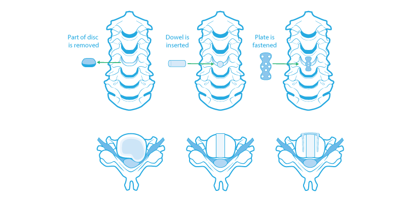
Anterior Cervical Diskectomy and Fusion

The cervical intervertebral disk is made up of a fibrous outer ring called the annulus and a gelatinous inner portion called the nucleus. The disk acts as a cushion between the vertebral bodies. When herniation occurs, the nucleus pushes through the annulus of the disk producing pressure on the nerve root, and/or spinal cord.
Nearly all patients complain of arm pain in the distribution of one or more nerve roots. Most have neck pain as well. Frequently, patients can be treated conservatively with success. However, when their ability to perform normal day to day functions is degraded for a substantial period of time (4-6 weeks) or in the presence of severe unrelenting pain or progressing neurological deficit, anterior cervical microdiskectomy with fusion is a highly effective treatment.
During microdiskectomy, the intervertebral disk and offending fragment are removed through a small, two to three inch incision in the crease of the neck. A dowel bone graft (from cadaver bone) or an intervertebral cage is inserted snuggly between the vertebral bodies to bring about a "fusion" or welding together of the two vertebral bodies. Other synthetic materials can be used to promote fusion as well. In many cases a plate is screwed into place to secure the bone graft. With success rates of around 70-80%, patients are able to return to work and full activity in an average of about 1-4 months. We have been performing anterior cervical diskectomy with interbody fusion on an outpatient basis for many years. It is well tolerated and preferred by patients over even a short hospital stay.
Your doctor will provide details of the procedure that is right for you as well as the benefits and risks. He will also provide instructions for your care before and after the procedure.
Posted on Mon, October 13, 2014
by Greg Neundorfer